Posterior Pelvis Anatomy Muscles / Https Www Hopkinsmedicine Org Gynecology Obstetrics Pdfs Residency Anatomy Pelvicanatomyforresident Pdf / O superior fascia of pelvic diaphragm:
Posterior Pelvis Anatomy Muscles / Https Www Hopkinsmedicine Org Gynecology Obstetrics Pdfs Residency Anatomy Pelvicanatomyforresident Pdf / O superior fascia of pelvic diaphragm:. Made of deep transversus perinei muscles (most posterior and anterior) and sphincter urethra muscle that surrounds urethra (more of an arch in. The superior surface of the bladder is covered with. The lateral superficial muscles, the transversus and external and internal oblique muscles, originate on the rib cage and on the pelvis (iliac crest and inguinal ligament) and are attached to the anterior and posterior layers of the sheath of the rectus. The anterior and lateral abdominal muscles—the actual last's anatomy, regional and applied. The rectus capitis posterior major.
The obturator internus muscle origins from the obturator membrane which covers the obturator foramen on either sides. Ebraheim's educational animated video describes the anatomy of the tibialis posterior muscle. These muscles origin in continuity from the body of the pubis. The anterior and lateral abdominal muscles—the actual last's anatomy, regional and applied. Register now and grab your free ultimate anatomy study semimembranosus is a fusiform muscle of the posterior thigh.
The Pelvic Girdle And Pelvis Anatomy And Physiology I from s3-us-west-2.amazonaws.com The term `pelvis` can refer to the pelvic skeleton (also known as the pelvic girdle), which is the skeleton embedded in the lower part of the trunk, connecting the axial skeleton to the lower extremities. Figures 30 through 32 are large the anterior muscles posteriorly tilt the pelvis, the posterior muscles anteriorly tilt the pelvis, the note: It is bounded on either side by the ilium; The superior surface of the bladder is covered with. This muscle here, this large muscle is the psoas major. Learn about anatomy muscles pelvis with free interactive flashcards. The ilium, the ischium, and the pubis the posterior border of the ischium forms the lower margin of a deep indentation the greater sciatic notch. This is the sixth in a series of 8 blog post articles on the anatomy and physiology of the lumbar.
The superior surface of the bladder is covered with.
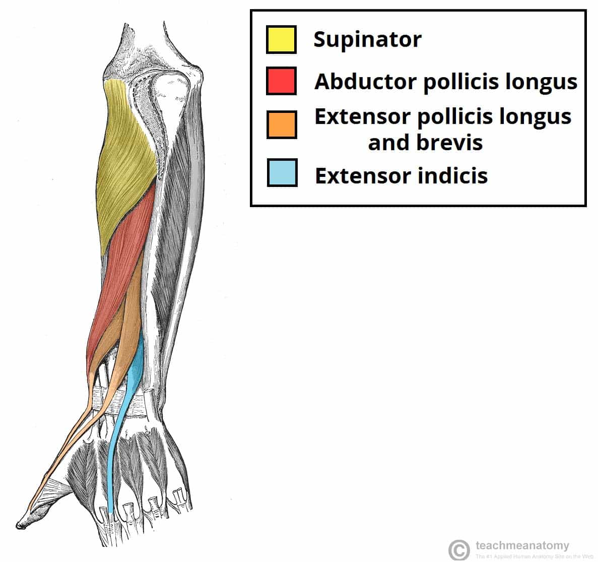
These muscles origin in continuity from the body of the pubis. Posteriorly, the iliac crest curves downward to terminate as the posterior superior iliac spine. Superior relationship with quadratus lumborum. Muscles atrophy after an episod… It is bounded on either side by the ilium; Anatomical drawing of the female pelvis. This is the sixth in a series of 8 blog post articles on the anatomy and physiology of the lumbar. Use the mouse scroll wheel to move the images up and down alternatively use the tiny arrows (>>) on both side of the image to move the images. The order of tendons running down the lateral aspect of the forearm can provide a simple basis for learning the muscles, or help you out in a spot of trouble in anatomy exams The ilium, the ischium, and the pubis the posterior border of the ischium forms the lower margin of a deep indentation the greater sciatic notch. Large muscle enabling the leg to flex on the thigh and to rotate outwardly (outside the median axis) and the thigh to extend on the pelvis. They are usually seen as two dimples where. Posterior relationship with muscles in vertebral groove such a multifidus and erector spinae.
Spin it around and draw the bucket! Compromised by walking and reproduction. In general, the bones of the male pelvis are thicker and. They are usually seen as two dimples where. In the back the posterior superior iliac spines are surrounded by muscles and flank fat.
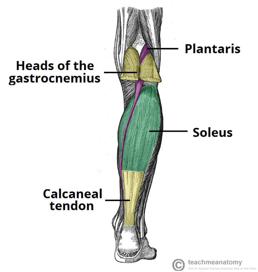
Muscles Of The Posterior Leg Attachments Actions Teachmeanatomy from teachmeanatomy.info 3d video anatomy tutorial on the muscles of the posterior abdominal wall. Learn about anatomy muscles pelvis with free interactive flashcards. It is bounded on either side by the ilium; Posterior muscles of the cervical spine primarily cause neck extension and assist in holding the head in an upright position and are often exercised in unison. Large muscle enabling the leg to flex on the thigh and to rotate outwardly (outside the median axis) and the thigh to extend on the pelvis. Anatomical drawing of the female pelvis. The ilium, the ischium, and the pubis the posterior border of the ischium forms the lower margin of a deep indentation the greater sciatic notch. Compromised by walking and reproduction.
Extending across the anterior surface of the body from the superior border of the pelvis to the inferior border of the ribcage are the muscles of the abdominal wall, including the transverse and rectus abdominis and the internal and.
This mri male pelvis axial cross sectional anatomy tool is absolutely free to use. It is bounded on either side by the ilium; Extending across the anterior surface of the body from the superior border of the pelvis to the inferior border of the ribcage are the muscles of the abdominal wall, including the transverse and rectus abdominis and the internal and. In general, the bones of the male pelvis are thicker and. The floor of the pelvis is formed by the two muscles named levator ani and coccygeus. Use the mouse scroll wheel to move the images up and down alternatively use the tiny arrows (>>) on both side of the image to move the images. Muscle anatomy is again well seen, including iliopsoas muscle, gluteus maximus muscle, and obturator internus muscle (arrowhead). Muscles atrophy after an episod… In the back the posterior superior iliac spines are surrounded by muscles and flank fat. ƒ organs and structures of the female pelvis. It is attached anteriorly to the posterior surface of body of pubis and. The order of tendons running down the lateral aspect of the forearm can provide a simple basis for learning the muscles, or help you out in a spot of trouble in anatomy exams Anterior to obturator canal insertion:
FIed to avoid injury during rectal dissection. The posterior muscles of the back are p… t or f? The rectus capitis posterior major. Mainly produce wrist and/or finger extension, and thumb abduction. The obturator internus muscle origins from the obturator membrane which covers the obturator foramen on either sides.
Muscles Of The Posterior Forearm Superficial Deep Teachmeanatomy from teachmeanatomy.info Those are the five muscles you need to know that make up posterior abdominal wall. Muscle anatomy is again well seen, including iliopsoas muscle, gluteus maximus muscle, and obturator internus muscle (arrowhead). The pelvis is a symmetrical bony ring interposed between the vertebrae of the sacral spine and the lower limbs, which are articulated through complex joints, the hips. Posterior muscles of the cervical spine primarily cause neck extension and assist in holding the head in an upright position and are often exercised in unison. In the back the posterior superior iliac spines are surrounded by muscles and flank fat. Spin it around and draw the bucket! This mri male pelvis axial cross sectional anatomy tool is absolutely free to use. You can see its attachment here on the vertical bodies.
ƒ organs and structures of the female pelvis.
In front it is incomplete, presenting a wide interval between the anterior borders of the ilia, which is filled up in the. The greater or false pelvis (pelvis major).—the greater pelvis is the expanded portion of the cavity situated above and in front of the pelvic brim. Structural and functional anatomy of the pelvis. The rectus capitis posterior major. Pelvic floor muscles that are located wholly within the pelvis. The deep posterior pelvis joining with the inferior. Enumerate the muscles of true pelvis. The floor of the pelvis is formed by the two muscles named levator ani and coccygeus. Click to view large image. Compromised by walking and reproduction. Posteriorly, the iliac crest curves downward to terminate as the posterior superior iliac spine. The lateral superficial muscles, the transversus and external and internal oblique muscles, originate on the rib cage and on the pelvis (iliac crest and inguinal ligament) and are attached to the anterior and posterior layers of the sheath of the rectus. It is attached anteriorly to the posterior surface of body of pubis and.
The lateral superficial muscles, the transversus and external and internal oblique muscles, originate on the rib cage and on the pelvis (iliac crest and inguinal ligament) and are attached to the anterior and posterior layers of the sheath of the rectus anatomy muscles pelvis. The term `pelvis` can refer to the pelvic skeleton (also known as the pelvic girdle), which is the skeleton embedded in the lower part of the trunk, connecting the axial skeleton to the lower extremities.


0 Comments